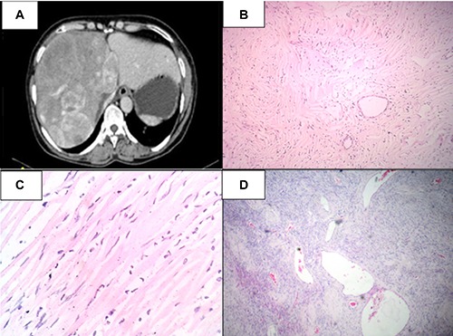
Figure 1.

A) Contrast enhanced computed tomography abdomen showing heterogeneously enhancing mass occupying right lobe and segment 4; B) microscopically, the tumor had low to moderate cellularity comprised of spindle to fibroblast-like cells in a collagenous background [hematoxylin and eosin (H&E), 40×]; C) the cells had spindle to elongated nuclei with variable indistinct eosinophilic cytoplasm (H&E, 100×); D) many thin walled ectatic blood vessels were interspersed within the tumor cells and resembled hemangiopericytoma-like areas (H&E, 40×).
