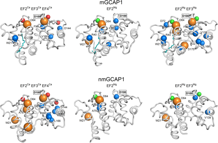
Figure 4. Highest degree hubs identified by PyInteraph in the PSN of mGCAP1 (top panels) and nmGCAP1 (bottom panels) in their EF2CaEF3CaEF4Ca (left), EF2Mg (center) and EF2MgEF3Mg (right) forms.
Secondary structure is represented in grey cartoons, Ca2+ ions are shown as red spheres, Mg2+ ions are shown as green spheres, the myristoyl group is represented as teal sticks, Cα of degree 7 hub residues are represented in blue spheres, Cα of degree 8 hub residues are represented in orange spheres with increased radius. Residues whose mutation is associated with cone, cone-rod or macular dystrophies are labelled in bold and framed.

