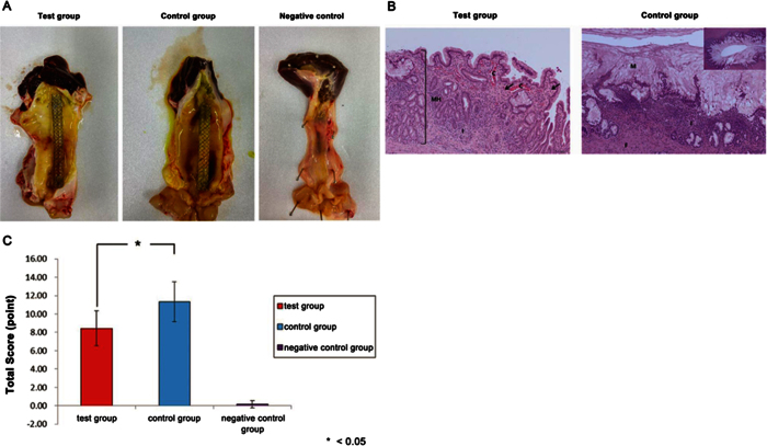
Figure 7. Pathological analysis.
(A) Gross findings. Compared with the negative control, the test tissue sample showed mild erythematous mucosal change and a normal SEMS configuration. In the control group, more severe erythematous change and bile-clogged membrane stent were noted. (B) Microscopic findings. The test group showed minimal mucosal hyperplasia (MH), necrosis (arrow), inflammation (I), and congestion (C) (left; HE stain, ×100). The control group showed remarkable mucosal hyperplasia with mucous secretion (M), fibrosis (F), and inflammation (I) (right; HE stain, ×100 and ×40 in the right upper panel). (C) Total pathology score was significantly lower in the test group.

