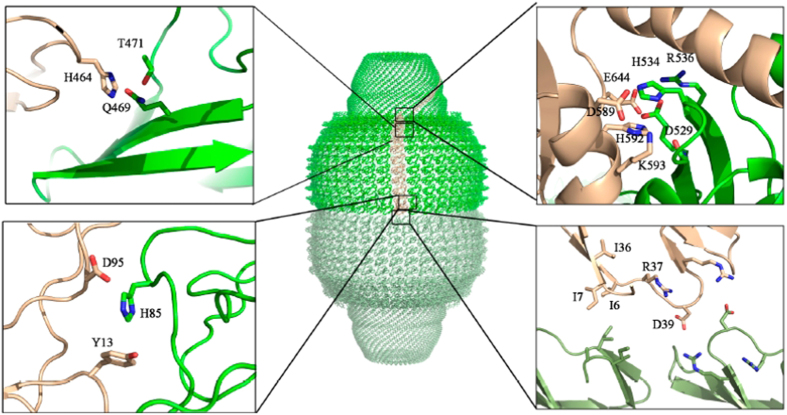
Figure 5.
(A) Structure of the vault shell, colored in green and pale green for the top and bottom half-vault moieties, respectively. One of the 78 MVP copies forming the particle shown in brown (PDB id: 4JL8)6. The left insets show a close up of the MVP-MVP lateral electrostatic interactions mediated by histidines in the R2 (bottom) and R9 (top) domains. The top right inset shows the MVM-MVP lateral electrostatic interactions in the shoulder domain. The right bottom inset shows the R1-R1 interactions established at the half-vault interface.

