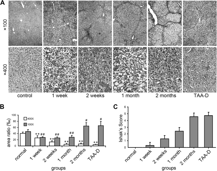
Figure 1. Change of reticular fibers and Ishak’s socre of TAA-treated rat liver tissues.
(A) Reticular fiber stain (Gomori’s reticular stain, ×100 and ×400 magnifications, for inserts, the corresponding background was erased); (B) Areas of reticular fibers, n = 6/group; (C) Ishak’s socre. ×400: *p < 0.05, **p < 0.01 vs. control group; ×100: #p < 0.05, ##p < 0.01 vs. control group.

