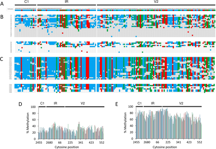
Figure 4. Bisulfite sequencing of TYLCSV DNA.
(A) TYLCSV fragment (938 nt) including portions of C1 and V1 coding sequences and the intergenic region (IR). (B,C) Cytosine methylation in individual clones from TYLCSV- (B) or TYLCSV/PSTVd-infected plants (C); the two blocks are from two experiments. Methylated cytosines (reported as C) are blue, red and green, if in the CHH, CHG or CG contexts, respectively. Un-methylated cytosines are reported as T. (D,E) Percentage of methylated cytosines within the TYLCSV amplicon in TYLCSV- (D) or TYLCSV/PSTVd-infected plants (E).

