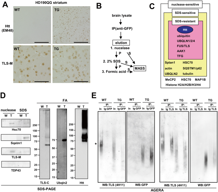
Figure 1. FUS/TLS binds to mutant huntingtin in vivo.
(A) Protein distribution of Htt-EGFP and FUS/TLS in HD190QG and control animals at 24 weeks. Scale bar: 50 μm. (B) Procedure of immunoisolation of mutant Htt aggregates from HD190QG brain. S: supernatant, P: pellet. (C) Summary of mass analysis using eluted fractions from GFP immunoprecipitates. (D) Western blot analysis of eluted fractions of GFP immunoprecipitates. (E) AGERA of immunoprecipitates of anti-GFP and anti-TLS-M eluted by 2% SDS and 1M DTT. Membranes were stained with anti-GFP or anti-TLS (4H11).

