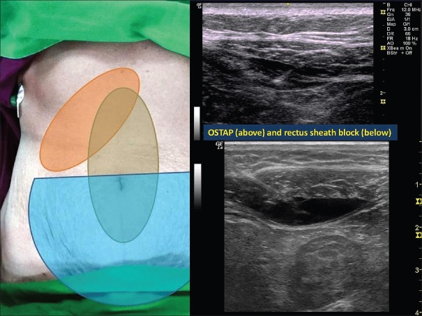
Figure 2.
Left: Comparison of sensory block achieved by bilateral rectus sheath block (grey area over midline), bilateral transversus abdominis plane block (green semi-circle over lower abdomen) and unilateral oblique subcostal transversus abdominis plane block (which can vary but approximately covers the area shaded in orange); Right: Above: Ultrasound image of oblique subcostal transversus abdominis plane block: the needle is seen depositing the drug (black area) between the posterior rectus sheath and the transversus abdominis muscle. Below: Rectus sheath block: the needle is seen depositing the drug between the rectus abdominis muscle and the posterior rectus sheath

