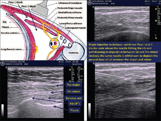
Figure 4.

Left above: Anatomical basis of pectoral nerves block. Left below: Initial ultrasound scan showing chest wall muscles, ribs and pleura. Right: Ultrasound images of performing combined Pecs-1 and Pecs-2 blocks in single injection technique

Left above: Anatomical basis of pectoral nerves block. Left below: Initial ultrasound scan showing chest wall muscles, ribs and pleura. Right: Ultrasound images of performing combined Pecs-1 and Pecs-2 blocks in single injection technique