Figure 1.
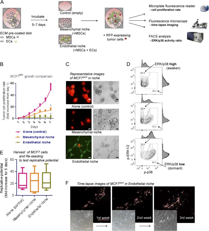
ER+ breast tumor cell MCF7 dormancy in bone marrow endothelial niche models. (A) Experimental scheme. BM MSCs and/or ECs were seeded on 96-well microplates and incubated until reaching confluence. For 3D cultures, Matrigel was coated (150 &L/cm2) onto the culture plate. Then, RFP-expressing tumor cells were sparsely seeded (200 cells/well) onto the niche cells or empty surfaces (control). Tumor cell proliferations were assessed by fluorescence intensity reader and microscope, and fluorescence-activated cell sorting (FACS). (B) Seven-day proliferation rates of MCF7 cells in niches (five sample sets per group; error bars: ± standard deviation (s.d.) *P < 0.05). (C) Representative images of tumor cells in the niches, captured after 7 days of co-culture (left: fluorescence (red and green); right: bright-field. scale bar = 100 &m). For visualization, ECs were prestained with CFSE (green). (D) FACS analysis of ERK1/2 and p38 MAR kinase activities of MCF7 tumor cells in niche. Upper: tumor cell alone; middle: tumor cell in mesenchymal niche; lower: tumor cell in endothelial niche. Blank arrows: ERK/p38 ratio-low (dormant) population; filled arrows: ERK/p38 ratio-high (awaken) population. (E) Replicative potentials of dormant MCF7 tumor cells. Tumor cells were harvested from niche and replated in 96-well microplates (500 cells/well). Box-plots represent fluorescence intensity fold-changes for 7 days (five samples per condition. horizontal bar = mean). (F) Time-lapse images of tumor cell dormancy in niche. MCF7RFP cells were cultured in endothelial niche for up to 21 days (fresh medium replaced every 48 h). Cells remain at almost the same number during the period. (Upper: fluorescence (red); lower: bright-field. scale bar = 500 &m). BM MSC, bone marrow-derived mesenchymal stem cell; EC, endothelial cell; ER, estrogen receptor; MAP, Mitogen-activated protein; p-ERK1/2, phosphorylated ERK1/2; p-p38, phosphorylated p38; RFP, red fluorescence protein. A full colour version of this figure is available at http://dx.doi.org/10.1530/ERC-16-0086.

 This work is licensed under a