Figure 3.
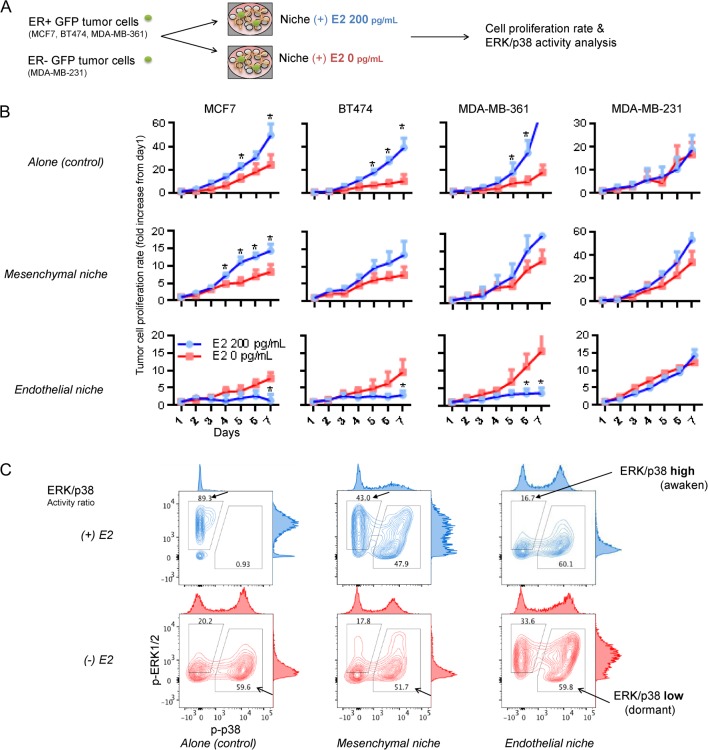
Increased ER+ tumor cell proliferation after estrogen depletion in bone marrow endothelial niche. (A) Experimental scheme. BM niche models were generated by co-culturing BM MSCs and/or ECs on ECM-coated microplates. GFP-expressing tumor cells were sparsely seeded (200 cells/well) onto the niche cells or empty surfaces (control). Tumor cell proliferations were compared in niches cultured using estrogen-supplemented (200 pg/mL) or un-supplemented (depleted) media. (B) Seven-day proliferation rates of tumor cells in niches. From left to right: ER+ MCF7, ER+ BT474, ER+ MDA-MB-361 and ER- MDA-MB-231. (Five sample sets per group; error bars: ±s.d. *P < 0.05). (C) FACS analysis of ERK1/2 and p38 MAR kinase activities of MCF7 tumor cells in estrogen-supplemented (blue) and estrogen-depleted (red) niches. Filled arrows: ERK/p38 ratio-high population. Blank arrows: ERK/p38 ratio-low population. A full colour version of this figure is available at http://dx.doi.org/10.1530/ERC-16-0086.

 This work is licensed under a