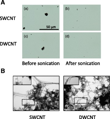
Fig. 1.

Optical microscope and TEM micrographs of CNT suspensions at 1.0 mg/ml concentration. a Optical microscope before and after dispersion by a cup-type sonicator at 100 W, 80 % pulse mode, for 10 min twice. b Transmission electron microscope (TEM) micrographs of SWCNTs and DWCNTs dispersed in the dispersion medium. The areas, which include well-dispersed CNTs, were highlighted within the dotted lines. SWCNT: single-walled carbon nanotube, DWCNTs: double-walled carbon nanotube
