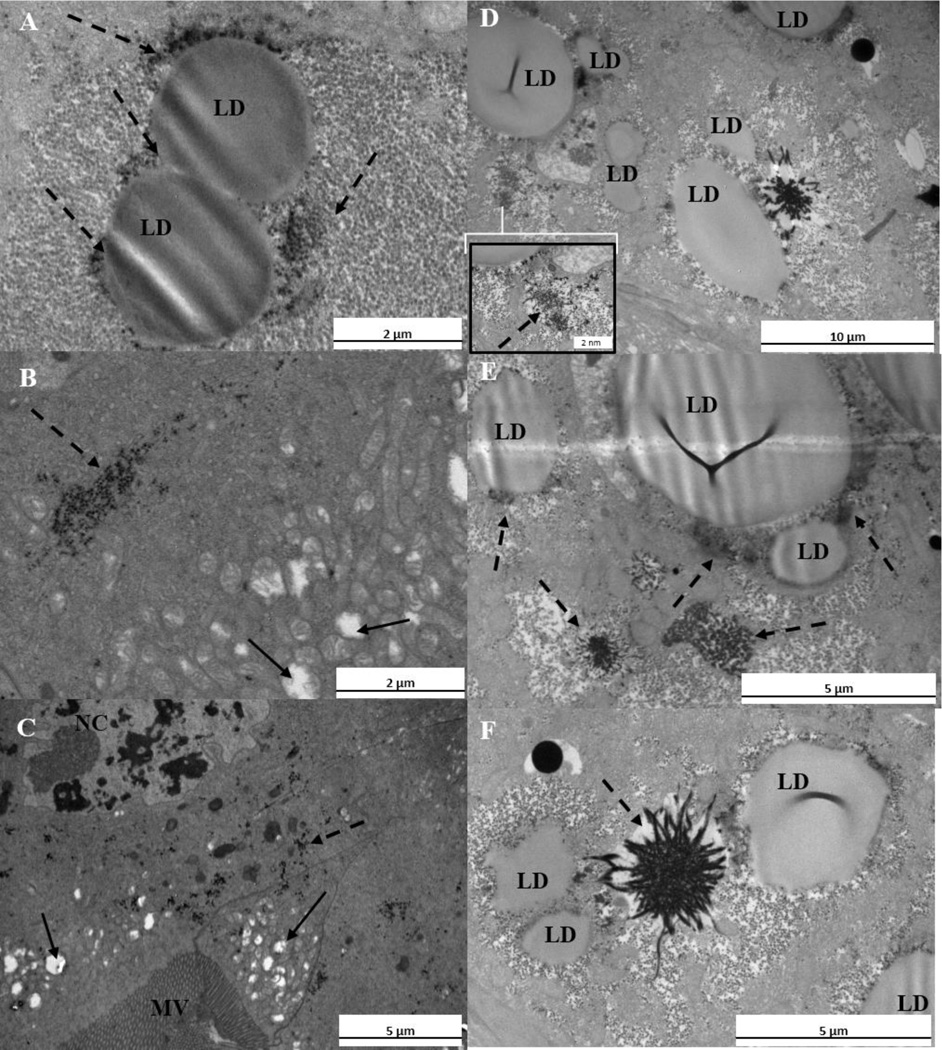
Fig. 2.
A–F. Transmission electron micrographs of C. riparius after 10 d of exposure in treatment A. The symbols MV, LD and NC indicate microvilli layer, lipid droplet and nucleus, respectively. Solid arrows indicate mitochondria, while dashed arrows indicate precipitates. (A) Lipid droplets and precipitates between them. (B) Precipitates in the tissue and swollen mitochondria (C) The structure of the nucleus, swollen mitochondria and precipitates next to the microvilli layer. (D) Lipid droplets and precipitates located near of them; in the inserted Figure is a higher magnification of the precipitate. (E) Large precipitates close to lipid droplets. (F) Lipid droplets and large precipitate between them.

