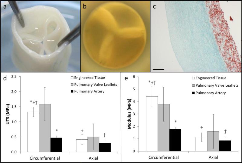
Figure 1.
Images of the tubular tissue-engineered valve (a) showing the leaflets, root, and (b) short axis view of the valve showing leaflet coaptation under physiologic backpressure. (c) Trichrome stained cross-section of the engineered tissue showing collagen (green) and non-collagen proteins (red); the scale bar is 250 μm. (d) Ultimate tensile strength and (e) tangent modulus of the engineered issue (n=4 for circumferential and axial), pulmonary valve leaflets (circumferential: n=3, axial: n=2), and the pulmonary artery (circumferential: n=4, axial: n=3) in the circumferential and axial directions at the time of implant. Paired symbols indicate a p-value < 0.05 between the two groups.

