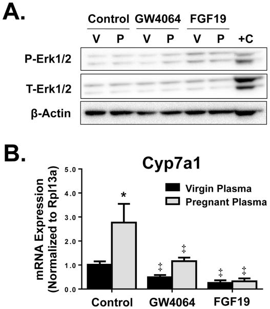
Fig. 6.
Cyp7a1 regulation in sandwich-cultured primary mouse hepatocytes. (A) Erk1/2 activation as indicated by phospho-Erk1/2 (P-Erk) compared to total Erk1/2 (T-Erk) after 1 hour treatment and (B) mRNA expression of Cyp7a1 after 24 hour treatment of sandwich-cultured primary mouse hepatocytes with media containing 20% pooled virgin or pregnant mouse plasma, in the presence of GW4064 (5 μM), recombinant FGF19 protein (20 μg/ml), or vehicle (DMSO). Western blots were performed using whole cell lysates. V, virgin plasma; P, pregnant plasma, mouse liver positive control. Data are presented as mean relative expression ± SD (n=3 donors/independent experiments in triplicate). Black bars represent virgin mouse plasma-treated hepatocytes and grey bars represent pregnant mouse plasma-treated hepatocytes. Asterisks (*) represent statistically significant difference (p≤0.05) compared with plasma-treated virgin controls. Double daggers (‡) represent statistically significant difference (p≤0.05) compared with plasma-treated pregnant controls.

