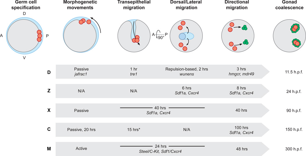
Figure 1. Shared themes in the migration path of embryonic germ cells.
Shown are highly stylized schematics of an embryo not meant to represent any one species. The ‘species-less’ embryo is shown at six key events during germ cell migration in chronological order from left to right. First, germ cells (red) are specified, often at the posterior or edge between embryonic (gray) and extra-embryonic (blue) tissue. Second, germ cells move during somatic morphogenetic movements (dashed arrow). In many species, germ cells move passively during gastrulation and often move within the developing mid or hindgut. Third, germ cells in several species undergo a transepithelial migration to exit the gut. Fourth, germ cells move dorsally and laterally to sort into two populations. Fifth, germ cells undergo a sustained, directed migration toward the developing somatic gonad (green circles). Sixth, germ and somatic gonadal cells coalesce to form the complete embryonic gonad. Shown underneath each stage of germ cell migration is a table with characteristic, key factors and length of stage noted for specific model organisms: D-Drosophila, Z- Zebrafish, X- Xenopus, C- Chicken, M- Mouse. Hpf – hours post fertilization. A- anterior, P- posterior, D- dorsal, V- ventral *Unlike other species, chicken germ cells migrate through the vascular epithelium rather than the gut epithelium.

