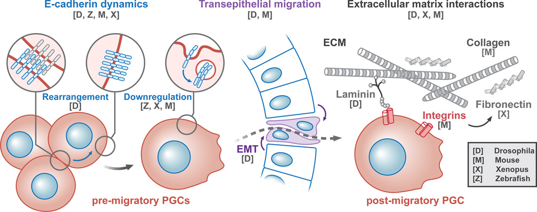
Figure 2. Transitions between migratory stages coincide with adhesion dynamics.
Shown is a stylized schematic of germ cell transepithelial migration that occurs during embryogenesis in many species. The transition between tissue types often accompanies changes in localization or downregulation of the cell-cell adhesion protein, E-Cadherin (blue links on red germ cells). In Drosophila, transepithelial migration requires an endodermal (blue columnar cell) epithelial to mesenchymal transition (EMT) (purple cells). Once out of the gut, germ cells in some species make contacts with select extracellular matrix (ECM) (gray crosshatch). The model organism for which specific factors and processes have been discovered is noted as a single letter with the key code in the bottom right corner.

