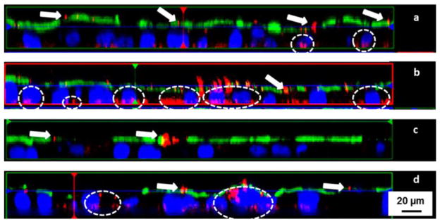
Figure 4.
Fluorescence microscopic z-scans of 200 nm fluorescent red polystyrene nanoparticles incubated with (a) Caco-2 monolayer, (b) Caco-2/M cell double culture, (c) Caco-2/HT29-MTX double culture, and (d) Caco-2/HT29-MTX/M cell triple culture. The arrows depict particle aggregates caught on the luminal side, and circles indicate nanoparticle uptake/penetration. The presence of mucus dramatically decreased nanoparticle uptake, while M cells facilitate nanoparticle translocation. Adapted from reference [111] with permission.

