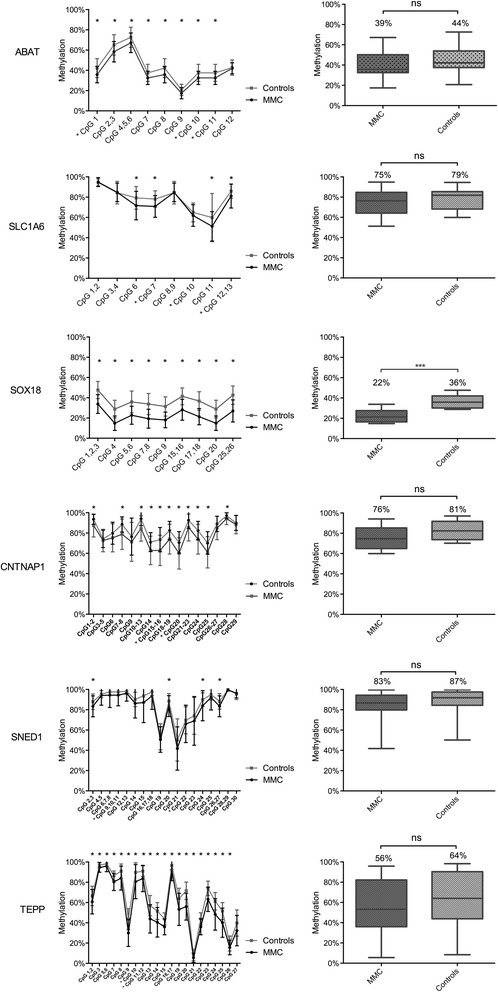
Fig. 2.

Validation of the top six differentially methylated genes by Sequenom EpiTYPER in MMC patients versus controls. Left: methylation pattern for each CpG unit within the amplicons. Multiple t test was performed for each CpG. *P value <0.05. Right: boxplot representing methylation pattern with box = 25th and 75th percentiles; bars = min and max values. The mean methylation level of each group is shown above the plot. The validation study is performed for 83 MMC patients and 30 controls. *CpGs were first identified by HM450k
