
Fig. 4.
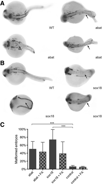
Phenotype analysis of gene overexpression in zebrafish embryos. Pax2a staining after microinjection of abat mRNA (a) and sox18 mRNA (b). Wild-type (WT) zebrafish show expression in the hindbrain, hindbrain-midbrain boundary, neural tube, mesoderm, optic stalk, otic vesicle, and pronephric duct. Spinal cord malformation is indicated with an arrow. c Phenotype analysis after pax2a staining at 24 hpf resulted in respectively 50 and 74 % embryos with an affected phenotype after abat and sox18 overexpression. Folic acid supplementation after gene overexpression did not significantly influence the phenotype. ***P value <0.001