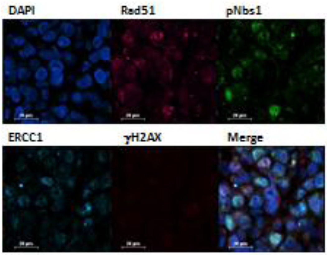
Figure 2. DNA damage multiplex assay utilized on an MX-1 BRCA-deficient breast cancer xenograft 24 hours after beginning treatment with irinotecan at 7.5 mg/kg.
False color assignments are as follows: Rad51 (pink), pS343-Nbs1 (green), ERCC1 (cyan), and γH2AX (red). A representative 40× image is shown. Cells expressing both pS343-Nbs1 (green) and γH2AX (red) are yellow on the merged image. Scale bar = 20 µm.

