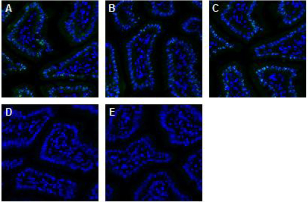
Figure 3. Peptide competition assay demonstrates pS343-Nbs1antibody specificity on the positive control tissue.
Representative images of formalin fixed, paraffin embedded mouse jejunum from serial cut slides. Tissue was stained with (A) 3 µg/ml of pS343-Nbs1 primary antibody, or pS343-Nbs1 protein incubated overnight with (B) peptide solution buffer, (C) 10 fold molar excess of the unphosphorylated peptide sequence or (D) 10 fold molar excess of the peptide sequence phosphorylated at S343. (E) A slide was also stained with a monoclonal rabbit isotype control. Images were extracted from a 20× Aperio scan with DAPI (blue) and pS343-Nbs1 (green) shown.

