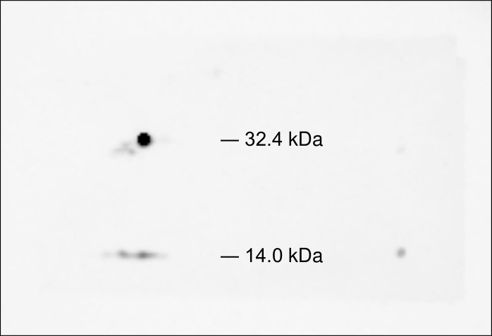
Fig 4. 2D western blot analysis using anti-TTR antibody.
The 32.4-kDa spot distinctly reacted with the anti-transthyretin antibody. Weak immunoreaction at 14.0 kDa was also detected and was interpreted as a monomer of TTR. The blotted membrane was stained with gold-colloid stain, and the area of immunoreaction was confirmed to correspond to the 32.4-kDa spot detected by 2D DIGE.

