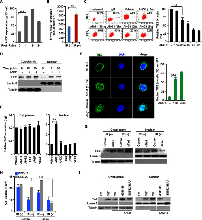
Fig. 2. ANG1 induces TIE2 nuclear localization.
(A) Increase of total ANG1 protein levels in U251.Tie2 cells in response to IR. (B) ANG1 protein levels of expression upon in vivo IR treatment of GSC-20–derived intracranial xenografts. HPF, high-power field. (C) Decrease of cell membrane–bound TIE2 upon ANG1 exposure in U251.Tie2 cultures. IgG, immunoglobulin G. (D) ANG1 exposure results on TIE2 nuclear translocation. (E) TIE2 localizes in the nucleus of human umbilical vein endothelial cells (HUVECs) upon ANG1 exposure, as assessed by immunofluorescence and confocal microscopic analysis. DAPI, 4′,6-diamidino-2-phenylindole. (F) TIE2 protein levels in cytoplasmic and nuclear U251.Tie2 cellular compartments after exposure to several ligands. bFGF, basic fibroblast growth factor; VEGF, vascular endothelial growth factor. (G and H) Soluble TIE2 (sTIE2) (G) jeopardizes IR-induced TIE2 nuclear translocation and (H) sensitizes GSCs to IR. (I) NLS mutations jeopardize TIE2 nuclear translocation upon ANG1 exposure. Data represent means ± SD; **P ≤ 0.01, ***P ≤ 0.001.

