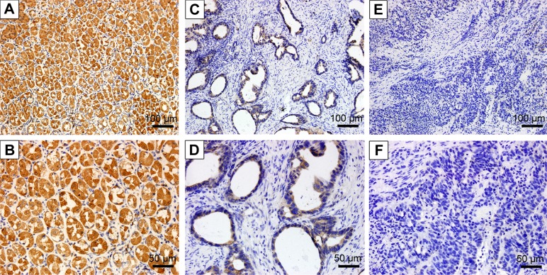
Figure 2.
Immunohistochemical analysis of ALDOB in GC and nontumor tissues.
Notes: Positive staining of ALDOB in nontumor gastric tissue: (A) 200×; (B) 400×. Positive staining of ALDOB in GC tissue: (C) 200×; (D) 400×. Negative staining of ALDOB in GC tissue: (E) 200×; (F) 400×.
Abbreviation: GC, gastric cancer.

