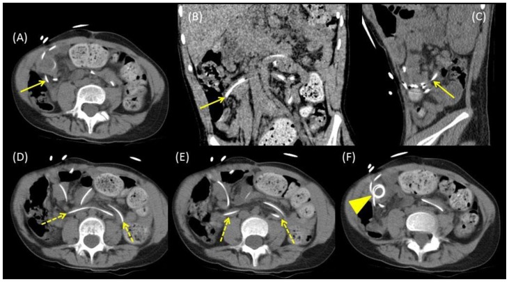
Figure 3.
8-year-old female with VP shunt erosion into the small bowel.
Findings: (A–C) CT of the abdomen and pelvis demonstrates the erosion site of the VP shunt catheter into the small bowel in axial (A), coronal (B), and sagittal (C) planes (arrows). (D and E) The VP shunt catheter is seen within several small bowel loops (dashed arrows) on axial CT images. (F) The VP shunt catheter is coiled outside of the bowel in the right lower quadrant (arrowhead), this likely corresponded to the central radiotracer organization in the abdomen seen on the VP shunt patency scan.
Technique: Axial CT of the abdomen/pelvis without contrast, 120kvp, 100 mA, 5 mm slice thickness. Coronal and sagittal thickness 1 mm.

