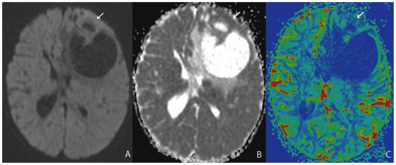
Figure 2.
13 month-old female infant with increasing head circumference and pathology proven DIG.
A) Findings: There is no no significant increased signal of the solid nodule (arrow) with respect to the normal brain parenchyma.
Technique: Axial DWI b=1000 image (1.5T, TR=7199, TE=97)
B) Findings: There is relative isointensity of solid tumoral ADC signal compared to adjacent brain parenchyma.
Technique: Axial ADC map image (1.5T, TR=7199, TE=97)
C) Findings: There is relative hypoperfusion of the solid nodule in comparison with adjacent and contralateral gray matter.
Technique: Axial rCBV map from DSC (1.5T, TR=1630, TE=49, contrast=Multihance 0.1mmol/kg, first pass bolus)

