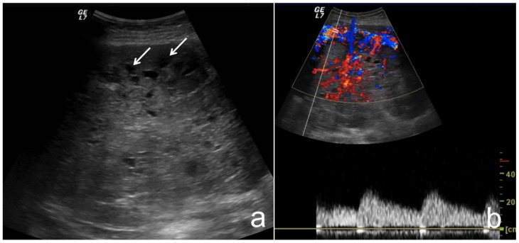
Figure 1.
37-year-old female with pancreatoblastoma.
a) Transverse transabdominal B-mode US image through the epigastrium shows a large heterogeneous mass, predominantly hypoechoic with small cystic cavities (white arrow). b) Transverse transabdominal Color Doppler US image shows that the mass has intense arterial and venous flow.
Technique: US (GE Logic 7 Pro), curved transducer, 3.5 MHz

