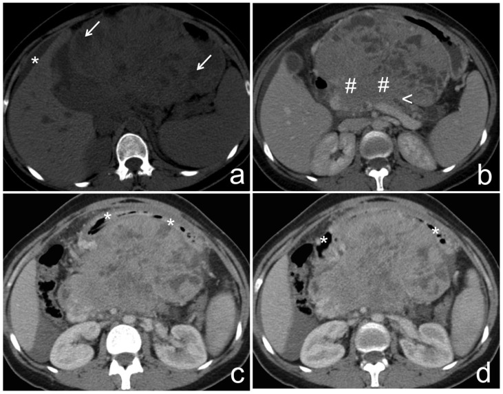
Figure 2.
37-year-old female with pancreatoblastoma.
a) Non-contrast-enhanced CT, axial reconstruction, shows a large intra-abdominal mass, centered at the epigastric region, presenting partially well-defined and lobulated contours, measuring 11,4×16,7×17,0 cm (antero-posterior, transverse and longitudinal axis, respectively). The mass is predominantly solid, with internal low-density cavities presumably cystic (white arrow). Note peri-hepatic ascites (asterisk).
b–d) Portal phase enhanced CT, axial reconstruction, shows that the solid component of the mass enhances, and is in continuity with the pancreatic body (cardinal). Note distal pancreatic ductal dilatation (arrowhead), suggesting its involvement. The mass pushed away the neighboring structures. The duodenal arch (asterisk) was anteriorly deviated, suggesting a retroperitoneal origin.
Technique: MCTD (Philips, Brilliance, 6), 120 kV, 155 mAs, 5 (a) and 3 mm slice thickness (b–d), 100 ml of iobitridol 350 mg/ml contrast media.

