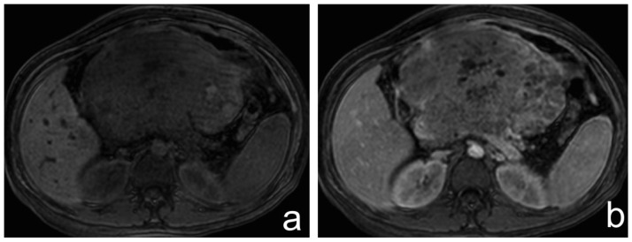
Figure 5.
37-year-old female with pancreatoblastoma.
a and b) Axial T1-weighted fat saturated MR image before and after gadolinium administration, respectively. The solid components show moderate enhancement.
Technique: 1,5 Tesla MRI (Philips Medical Systems Achieva). Image a: TR 3,6, TE 1,8, 5 mm slice thickness, non-contrast. Image b: TR 3,6, TE 1,8, 5 mm slice thickness following 5 ml of gadobutrol 1 mmol/ml, late arterial phase.

