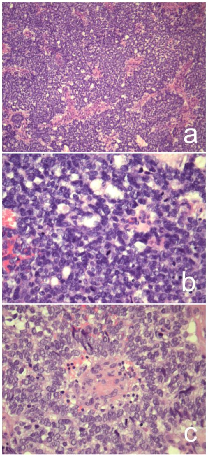
Figure 6.
37-year-old female with pancreatoblastoma.
Microscopic pathology of haematoxylin-eosin staining, original magnification ×100 (a) and ×400 (b–c). Microscopy showed abundant small tumor cells that exhibited an organoid growth pattern with focal acinar features. There are a few squamoid corpuscles.

