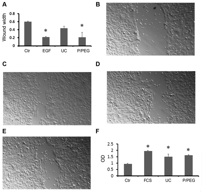
Figure 5.
Biological activity of extracellular vesicles (EVs) purified by ultracentrifugation and P/PEG precipitation from saliva and human liver stem cells (HLSCs). (A–E) Evaluation of wound healing on normal dermal keratinocytes (HaCaT) by scratch test. Quantitative evaluation of wound size reduction after 36-h incubation with the vehicle alone (Ctr), 10 ng/ml EGF as a positive control, and EVs isolated by ultracentrifugation (UC) or P/PEG precipitation (50,000 EVs/cell). Data are mean ± 1SD of three independent experiments evaluated in triplicate. ANOVA with Dunnet's multicomparison test was performed in all the samples vs. UC; *P<0.05. Representative micrographs of Ctr (B), EGF (C), UC EVs (D) and P/PEG EVs (E) induced wound healing. Original magnification, ×100. (F) Proliferation of TEC evaluated by BrdU incorporation after 12-h incubation with EVs isolated by UC or P/PEG precipitation (10,000 EVs/cell). As negative control, TEC was incubated with the vehicle alone in the absence of fetal calf serum (FCS); as positive control, cells were incubated with 10% FCS. Data are mean ± 1SD of three independent experiments evaluated in triplicate. ANOVA with Dunnet's multicomparison test was performed in all the samples versus Ctr; *P<0.05.

