. 2016 Sep 22;5:e17716. doi: 10.7554/eLife.17716

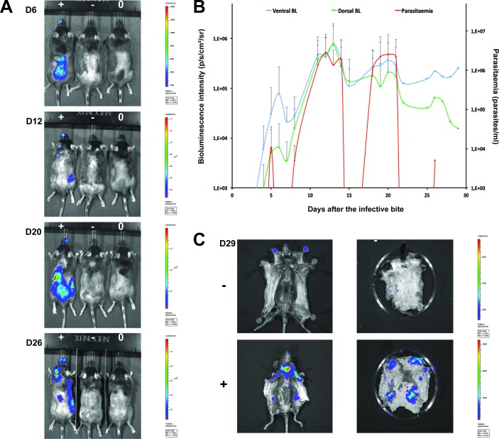
Figure 3. Dynamics of parasite distribution in the extravascular tissue of the skin and in the blood during a representative course of infection following natural transmission.

A total of seven mice were infected by the single infective bite of an individual G.m. morsitans on the belly with the T.b. brucei AnTat1.1E AMLuc/tdTomato strain. Panels A and C depict representative patterns. (A) Examples of bioluminescence profiles of 3 mice (+ bitten by an infected fly, - bitten by an uninfected fly and 0 not bitten) 6, 12, 20 and 26 days after the bite are shown. (B) Ventral (blue) and dorsal (green) bioluminescence (BL) intensities (in p/s/cm2/sr on the left Y-axis) and parasitaemia (in parasites/ml in red on the right Y-axis) were measured daily for 29 days and plotted as mean ± SD (n = 7 mice). (C) The entire skins of mice (+) and (-) were dissected for bioluminescence imaging 29 days after the bite. For the mouse (+), Figure 3—figure supplement 1 shows the bioluminescence profile of dissected organs, Figure 3—figure supplement 2 presents the skin inflammation, and Figure 3—figure supplement 3 shows labelled parasites in skin sections.

DOI: http://dx.doi.org/10.7554/eLife.17716.007

Figure 3.

Figure 3—figure supplement 1. Bioluminescence mostly originates from parasites in the skin.
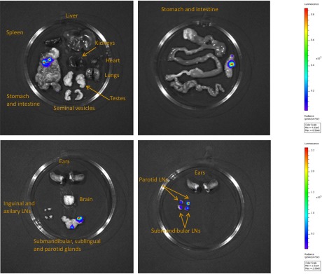
Figure 3—figure supplement 1.

Mouse (+) was sacrificed and dissected for bioluminescence imaging 29 days after the infective bite. Figure 3C shows the bioluminescence profile of its entire skin and dissected organs are shown here.

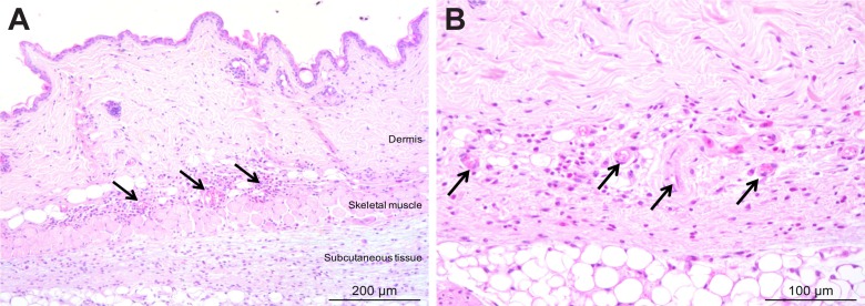
Figure 3—figure supplement 2. Mild inflammation of skin tissues one month after an infection by natural transmission.

Figure 3—figure supplement 2.

After 29 days, the most bioluminescent skin region of mouse (+) was dissected, fixed in paraformaldehyde, embedded in paraffin and stained with HE. Multifocal inflammatory infiltrates containing neutrophils were located in the dermis and subcutaneous tissue and associated with oedema (arrows in A). Inflammatory foci were generally centred on blood vessels (arrows in B).

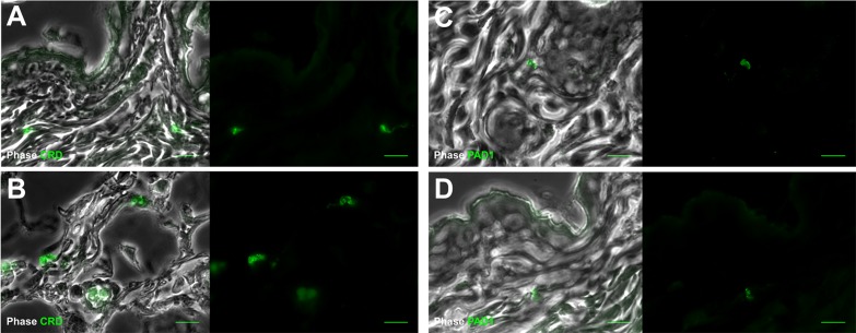
Figure 3—figure supplement 3. Extravascular parasites in the skin express both VSGs and PAD1 surface markers.

Figure 3—figure supplement 3.

After 29 days, the most bioluminescent skin region of mouse (+) was dissected, fixed in paraformaldehyde, embedded in paraffin and treated for IFA with the anti-CRD antibody that predominately labels parasites expressing VSGs (AB), or the anti-PAD1 antibody specific to transmission form 'stumpy' cells (CD).