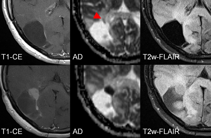
Fig 5. Female patient (41y) with a malignant transformation (anaplastic astrocytoma WHO III) of a primary resected fibrillary Astrocytoma WHO II in the right occipital lobe.
In the first examination (upper line) a subtle diffusion restriction is observed (red triangles) that predicts the future growth (lower line) of the malignant transformation, indicated by a hypointense cluster in the AD map. The difference between the first examination and the follow-up is 5 ½ months.

