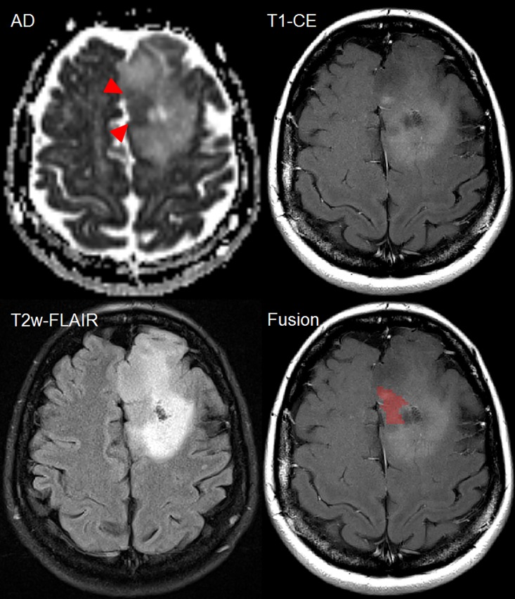
Fig 7. Female patient (44y) with malignant transformation of a primary resected oligoastrocytoma WHO II into anaplastic oligoastrocytoma III in the left frontal lobe demonstrating heterogeneity within the patchy contrast-enhancing region.
Increased diffusion restriction (red triangles) is noted at the same timepoint as new CE. Within the patchy area of CE, the spatial position of hypercellularity is visualized as red cluster (last image). Comparing T2w-FLAIR and axial diffusivity, the hypercellularity is found to be located in a T2w-hypointense region with a punctual CE focus. However, large proportions of patchy CE do not match with the diffusion restriction. Such information is important to consider for guidance of stereotactic biopsy and focal treatment planning.

