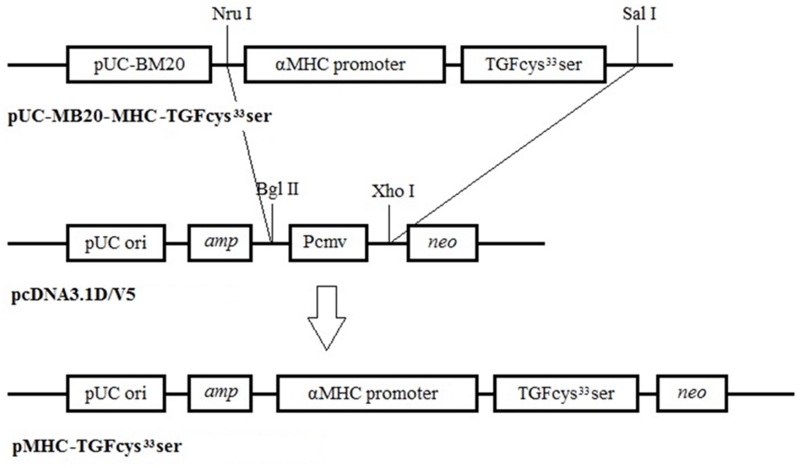
Figure 1.
pMHC-TGF-β1cys33ser knock-in vector construction. The pcDNA3.1D V5 vector was linearized by digestion with Bgl II, treated with Klenow to blunt the ends, followed by cutting with Xho I; these treatments resulted in the linearized pcDNA3.1D V5 vector bearing a blunt end at the Bgl II cut site and a sticky end generated by Xho I digestion. The MHC-TGF-β1cys33ser cassette was liberated from vector pUC-BM20-MHC-TGF-β1 by double digestions with Nru I and Sal I. As Nru digestion generates blunt ends and Sal I digestion generates a compatible sticky end to the sticky end generated by Xho I, the MHC-TGF-β1cys33ser cassette was directly subcloned into the pcDNA3.1DV5 vector prepared above to obtain the pMHC-TGF-β1cys33ser knock-in vector.

