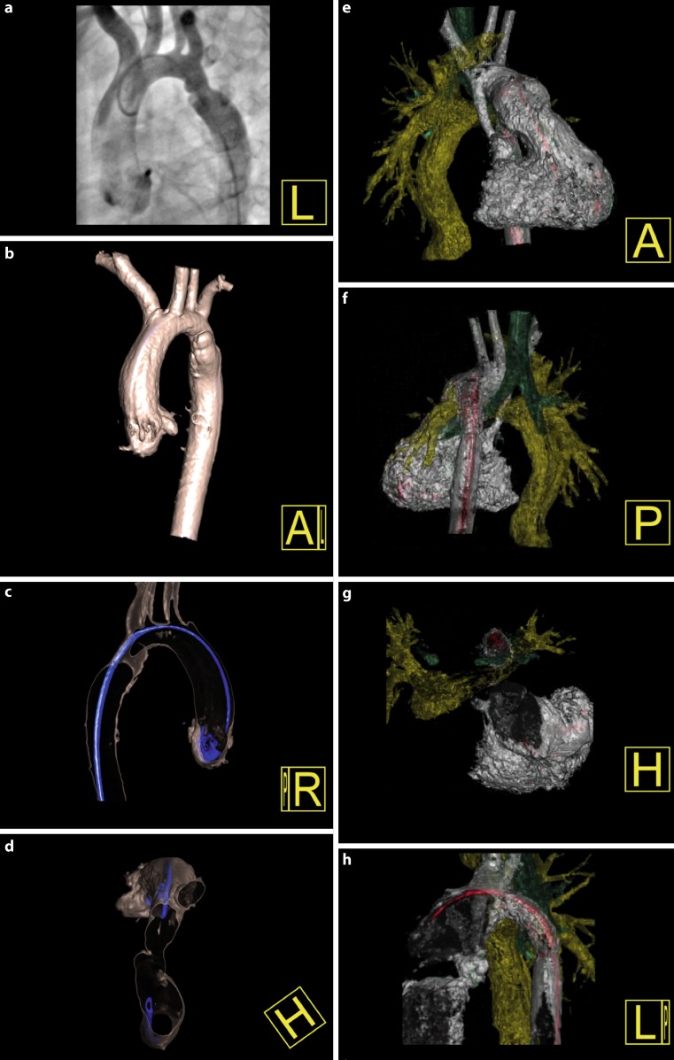
Fig. 3.
Displaying other vascular and extravascular structures with 3DRA. The left side displays a 2.5-year-old patient with a recurrent coarctation. 3DRA displayed a dissection on the anterior (b), lateral (c) and superior view (d), which was not clearly visible on the CA (a), leading to the decision of a stent implantation. The right side displays a 3.5-year-old patient with a recoarctation and a univentricular heart. 3DRA displayed an important interaction between the coarctation stent (white), the left pulmonary artery stent (yellow) and the left bronchus (green), which can be visualised from multiple angles (e-h)

