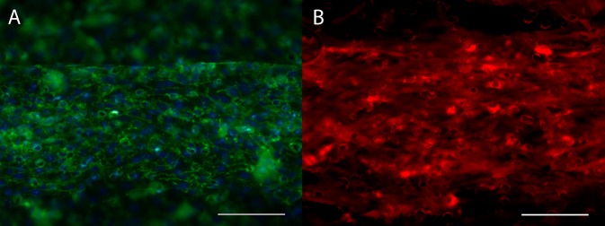
FIG. 4.
ECs formed a monolayer on the membrane in the EC channel of the device. (a) CD31 staining (green) with DAPI counterstain (cyan) demonstrates a confluent coverage and evidence of cell alignment in the direction of flow (left to right). (b) von Willebrand expression in ECs. Scale is 200 μm for the entire figure.

