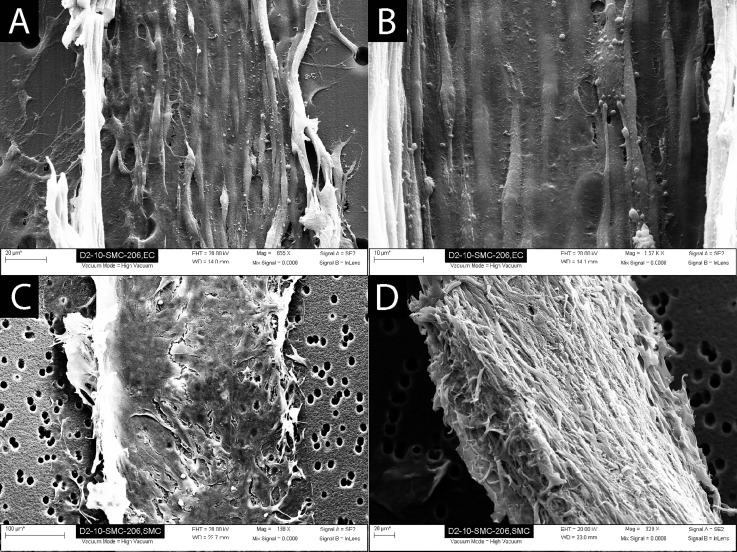
FIG. 5.
Scanning electron microscopy analysis of the devices demonstrated a confluent coverage of the endothelial channel side of the membrane with ECs, and confluent coverage of the SMC channel side of the membrane with SMCs. (a) EC layer (edges of cell layer peeled up during SEM sample preparation). (b) Higher magnification of EC layer demonstrating uniform EC coverage and alignment. (c) SMCs attached to the porous membrane. (d) SMCs removed from the membrane to demonstrate thickness of the cell layer (∼30 μm) and cell morphology.

