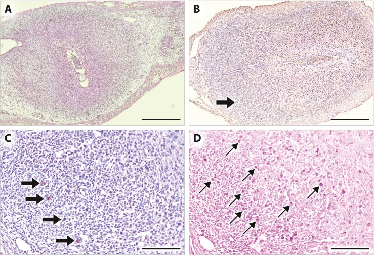
Fig. 2.

Distribution of uNK cells at early gestational stage in mice. Uterine sections from C57BL/6 female mice at gd 5.5 (a) and gd 6.5 (b−d) were stained with DBA lectin (A−C) or PAS (D). a At gd 5.5, DBA+ NK cells are scarcely detected. b At gd 6.5, DBA+ NK cells are increasingly detected in the mesometrial region. c Higher magnification of b. d Higher number of PAS+ NK cells are detected in the continuous section of c. Thin arrows: PAS+ cells, thick arrows: DBA+ cells. Bars, a, b 500 μm, c, d 100 μm
