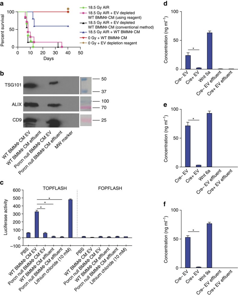
Figure 5. Presence of EV-packaged WNT in BMMΦ CM is critical for radio-mitigating function.
(a) Kaplan–Meier survival analysis of WT mice (n=10 per group) receiving CM/EV-depleted CM (500 μl per mice i.v.) derived from WT BMMΦ at 1 h and 48 h post 18.5 Gy AIR. Mice receiving WT BMMΦ CM showed 60% survival beyond 30 days compared with mice receiving EV-depleted WT BMMΦ CM (using reagent (P<0.004) or conventional method (P<0.002)) or untreated mice (P<0.0009; Log-rank (Mantel–Cox) test) where 80–100% of mice died within 12 days after irradiation (n=10 per group). Reagent (500 μl per mice i.v.) used for chemical depletion of exosome did not confer any toxicity to normal mice. (b) Immunoblot to detect exosomal markers TSG101, ALIX and CD9 in EV from WT BMMΦ CM or Porcn-null BMMΦ CM or respective effluents. EV from WT BMMΦ CM and from Porcn-null BMMΦ CM showed the presence of EV markers. However, EV markers were not detected in effluents. (c) TCF/LEF reporter assay. HEK293 cells having TCF/LEF luciferase reporter construct were treated with EV (prepared with the conventional method) from WT or Porcn-null BMMΦ CM or effluents or LiCl. Treatment with EV (100 μg ml−1) from WT BMMΦ CM showed higher Luciferase activity compared with EV (100 μg ml−1) from Porcn-null BMMΦ CM (P<0.0002) and effluent from WT BMMΦ CM or Porcn-null BMMΦ CM (P<0.0001 and P<0.0002 respectively; unpaired t-test, two-tailed). (d–f) ELISA to detect WNT5a, 6 and 9a in EVs from WT or Porcn-null BMMΦ CM and effluents. Presence of WNT5a, WNT6 and WNT9a were detected in EVs from WT BMMΦ CM (Cre− EV) but not in EVs from Porcn-null BMMΦ CM (Cre+ EV; P<0.0002; P<2.78E−05 and P<3.26E−06 respectively; unpaired t-test, two-tailed). WNT5a, WNT6 and WNT9a were also absent in effluents derived from WT or Porcn-null BMMΦ CM. Recombinant WNT5a, WNT6 and WNT9a were used as positive control respectively.

