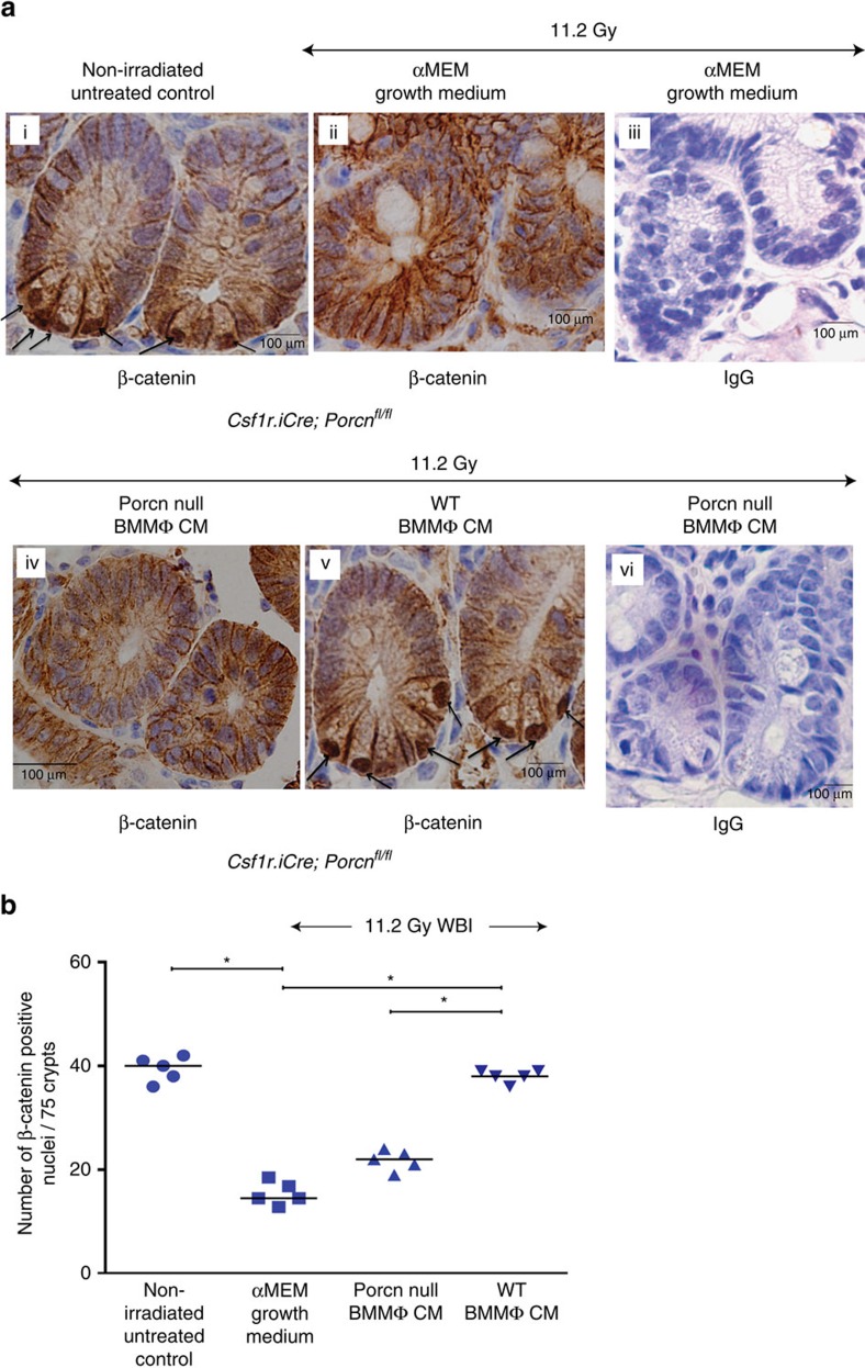
Figure 6. Macrophage-derived WNTs induce β-catenin activity in irradiated crypts.
(a) Representative microscopic images (× 60 magnification) of jejunal sections immunostained with anti β-catenin antibody to determine β-catenin nuclear localization in Csf1r.iCre;Porcnfl/fl mice. Nucleus stained with haematoxylin. Irradiated Csf1r.iCre;Porcnfl/fl mice receiving WT BMMΦ CM (i.v.) showed more nuclear β-catenin staining (dark brown; indicated with arrows) at the base of the crypt compared with mice receiving Porcn-null BMMΦ CM (i.v.) or αMEM growth medium (ii; nucleus stained blue). Fig iii and vi are representative IgG controls indicating lack of staining and thus showing specificity for the anti β-catenin antibody. (b) Nuclear β-catenin count: each data point is the average of the number of β-catenin-positive nucleus from 15 crypts per field, 5 fields per mice. Number of β-catenin-positive nucleus in irradiated Csf1r.iCre;Porcnfl/fl mice receiving WT BMMΦ CM is higher compared with Porcn-null BMMΦ CM (*P<1E−04 ) or αMEM growth medium (*P<1E−04). Treatment with Porcn-null BMMΦ CM and αMEM growth medium following irradiation showed significantly fewer β-catenin-positive nuclei than the non-irradiated control (*P<1.2E−04 and *P<1E−04 respectively; unpaired t-test, two-tailed).

