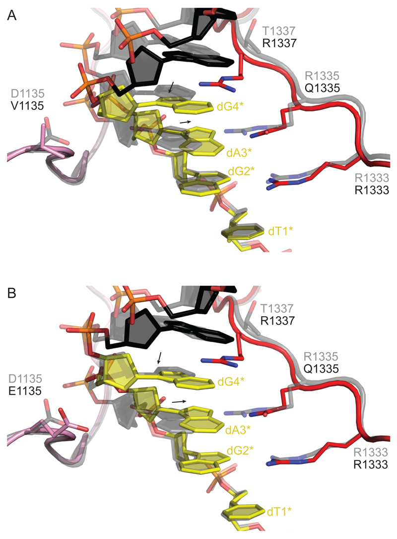
Figure 2. The VQR and EQR Variants Induce a Structural Distortion in the PAM DNA.
(A) Close-up view of the superimposed structures of the VQR variant with WT SpCas9. The PAM-interacting regions of the VQR Cas9 variant (colored pink and red) are not perturbed (Figure S2). The non-target DNA strand is colored black with the PAM highlighted in yellow. The superimposed WT SpCas9 structure is shown in gray. Arrows indicate shifts of the base and deoxyribose moieties of dA3* and dG4* in the VQR variant relative to the WT SpCas9 structure.
(B) Close-up view of the EQR variant superimposed with WT SpCas9. Color coding is as in (A)

