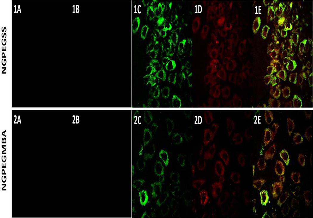
Figure 1. Confocal microscopy of primary bovine chondrocytes incubated at 37°C with (1) NGPEGMBA and (2) NGPEGSS.
(A) non-fluorescent nanoparticles as negative controls; (B&C) fluorescent nanoparticles after (B) 4 h and (C) 24 h incubation; (D) Lysotracker DND-99; (E) green nanoparticle overlay on red endolysosomes.

