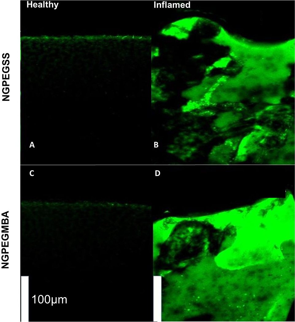
Figure 3. Mid-sagittal cross-sections of bovine knee explants from the load-bearing region of the femoral condyles with the top representing the articular surface after 24 h incubation with FITC-labeled nanoparticles.
(Top) NGPEGSSF and (Bottom) NGPEGMBAF. Left panel represents normal healthy cartilage incubated with fluorescent (A) NGPEGSSF or (C) NGPEGMBAF showing minimal diffusion of fluorescent particles. Right panel represents (B) NGPEGSSF and (D) NGPEGMBAF diffusing through the inflamed, aggrecan-depleted ex vivo cartilage explants. Scale bar represents 100 µm.

