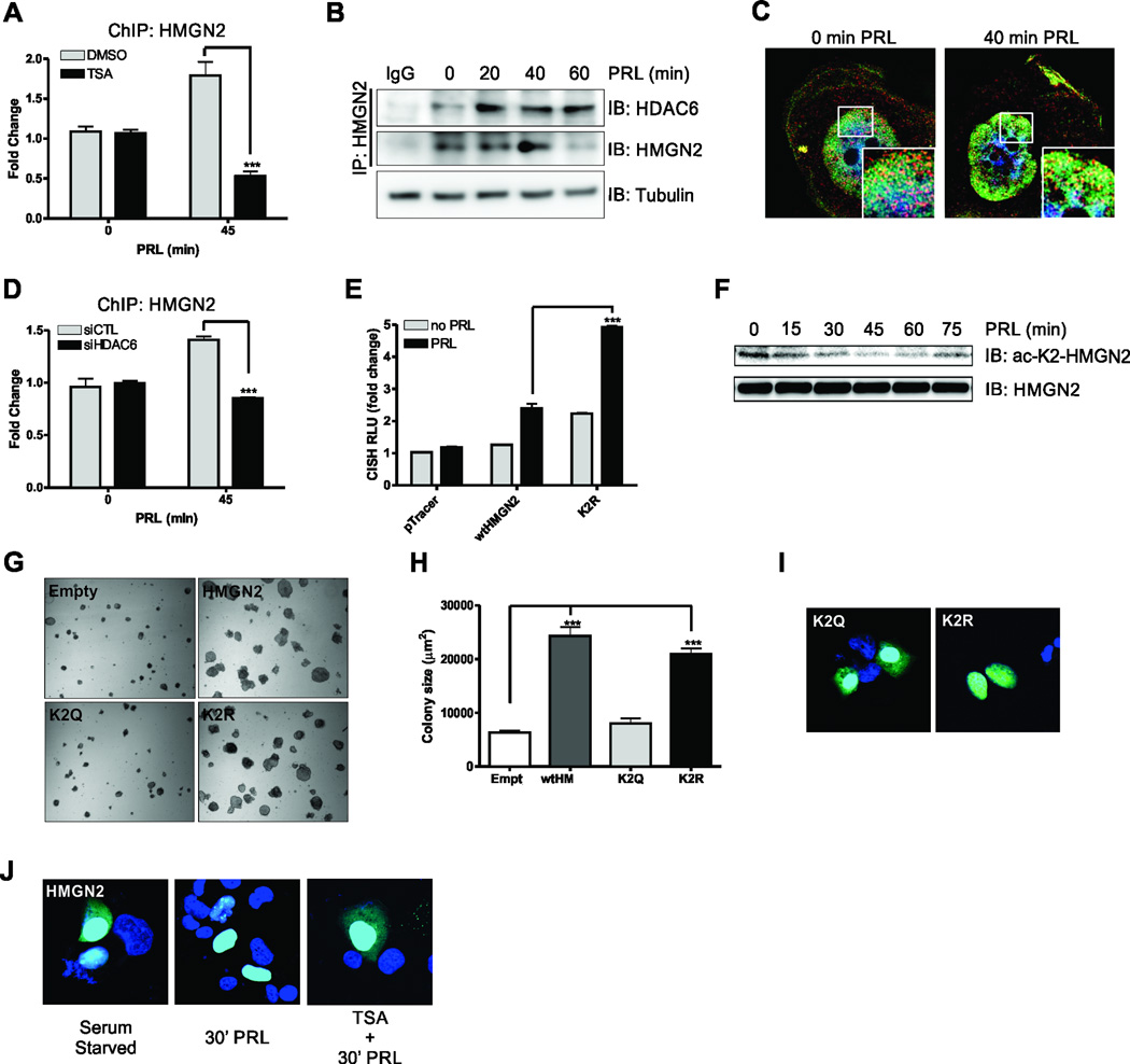
Figure 2. HMGN2 K2 deacetylation is critical for PRL-mediated signaling and the oncogenic potential of breast cancer cells.
(A) ChIP analysis of HMGN2 on the endogenous CISH promoter in MCF-7 cells treated with DMSO or TSA. Cells were serum starved for 24h and treated with 200nM TSA for 4h before stimulation with 250ng/mL PRL. Data are presented as mean ± SEM and were normalized to 18S RNA from 5% input. The fold change was determined using control transfected, no PRL as control. (B) CoIP of endogenous HMGN2 with HDAC6. MCF-7 ells were serum starved for 24h before treatment with PRL for the indicated timepoints. Tubulin immunoblot on 5% input was used as a loading control. (C) Super resolution microscopy of HDAC6 (red) and HMGN2 (green) using the Nikon N-SIM. T47D cells were serum starved for 24h before treatment with PRL for indicated timepoints. (D) ChIP analysis of HMGN2 on the endogenous CISH promoter in control or HDAC6 siRNA transfected MCF-7 cells. Cells were serum starved for 24h and treated with 250ng/mL PRL. (E) CISH luciferase assay in MFC-7 cells transfected with the control vector, HMGN2, or the deacetylated-K2 HMGN2 mimic (K2R). Cells were serum starved for 24h and treated with 250ng/mL PRL for 24h. (F) MCF-7 cells were serum starved for 24h and treated with 250ng/mL PRL for the indicated timepoints and immunoblotted with an α-acetyl-K2-HMGN2 antibody. Total HMGN2 was used as a loading control. (G) Soft agar colony formation of MCF-7 cells that had been transfected with control vector, HMGN2, the acetylated K2 HMGN2 mimic (K2Q), or the K2R deacetylated-K2 HMGN2 mimic. Representative images of colony growth and quantification of colony size (H) are shown. (I) Confocal microscopy of eGFP K2Q and K2R HMGN2 fusion constructs. T47D cells were transfected with eGFP fusion constructs to assess the localization of the HMGN2 mutants. (J) Confocal microscopy of T47D cells that had been transfected with eGFP HMGN2 fusion construct. T47D cells were serum starved for 24h and were then pretreated for 4h with DMSO or TSA before stimulation with 250ng/mL PRL for 30 min. All experiments were performed at least 3 times, unless otherwise noted. One-way or Two-way ANOVA with Bonferroini multiple comparisons test was used for statistical analysis where appropriate, * p<0.05; ** p<0.01; *** p<0.001.

