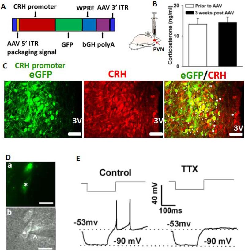
Figure 1. Identification of PVN-CRH neurons.
(A): Construct of AAV vector containing an eGFP gene under control of the rat CRH promoter. (B): Summary data show no significant change in circulating CORT levels prior to and 3 weeks after AAV-CRH vector injection (n = 7 rats, P > 0.05, paired t test). (C): Immunostaining depicts eGFP-tagged neurons as CRH immunopositive. The arrowheads indicate neurons with both eGFP and CRH immunoreactivity. The arrows indicate CRH-positive neurons without eGFP immunoreactivity. 3V, third ventricle. (D): eGFP-tagged PVN neurons (*) with an attached recording electrode (^) viewed with fluorescence illumination (a) and infrared differential interference contrast optics (b) in the brain slice. (E): Electrophysiological recordings showed that an eGFP-tagged neuron did not generate LTSs in response to depolarizing current pulses (30–45 pA) from a membrane potential of −90 mV in the absence and presence of 1 μM tetrodotoxin (TTX). Scale bars indicate 50 μm in C and 20 μm in D.

