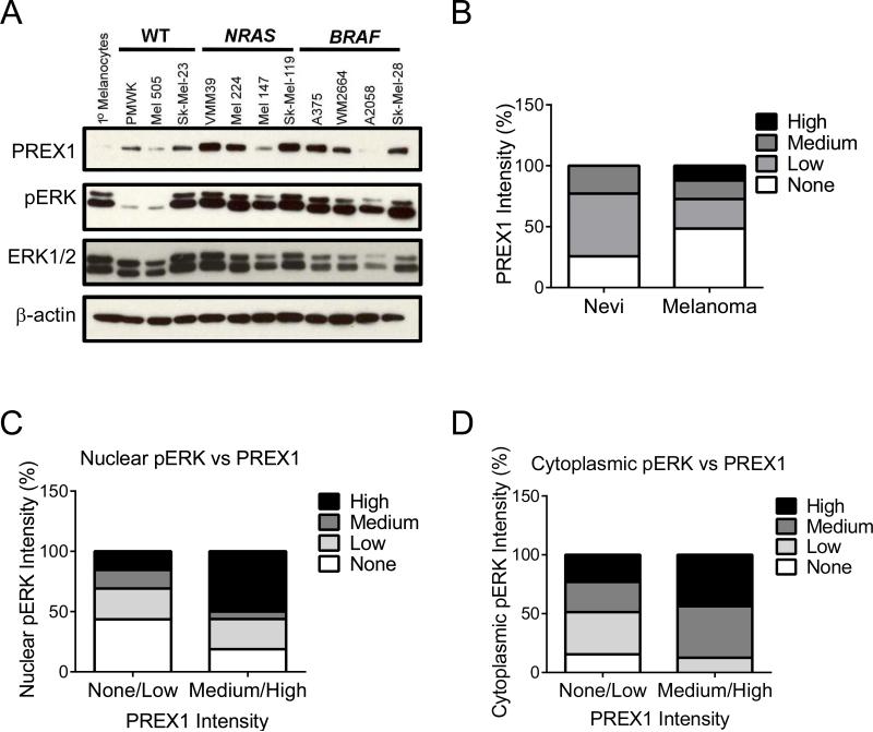
Figure 1. PREX1 protein levels are elevated in melanoma patient tumor tissues and cell lines, along with phospho-ERK.
(A) Western blot analysis of PREX1 protein, phospho-ERK (pERK) and total ERK1/2 in a panel of WT, BRAF- or NRAS-mutant human melanoma tumor cell lines. (B-D) Human tissue samples of benign melanocytic nevi and malignant skin cutaneous melanoma were subjected to IHC for PREX1 and pERK. Shown are (B) the distribution of PREX1 expression in nevi versus melanoma samples as measured by IHC; n=35 and 33, respectively. Samples were first binned according to no, low, medium or high staining intensity for each protein, and then the distribution was graphed to show the relationship between PREX1 and the percent of samples that stained positive for (C) nuclear pERK or (D) cytoplasmic pERK.

