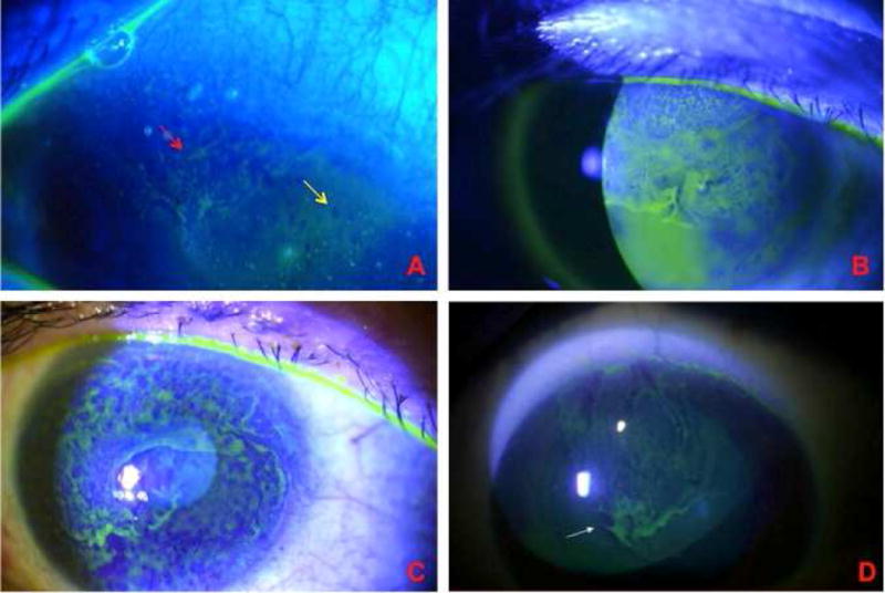
Figure 1. Contact lens-induced limbal stem cell deficiency.

A. Early involvement of the superior cornea. There is whorl-like epithelium (red arrow) adjacent to an area of punctate staining (yellow area), which is the earliest sign of LSCD. B Superior involvement of the cornea, characteristic of moderate CL-induced LSCD. C. Late-staining fluorescein diffusely in a whorled pattern as a confluent sheet across the cornea in late-stage CL-induced LSCD. D. Characteristic superior involvement of corneal epithelium.
