Fig. 5.
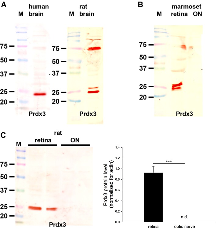
Western blot analysis of Prdx3 expression in brain, retina and optic nerve. Molecular weight markers (M, kDa) were used to determine size of detected gel products. a In tissue extracts from human and rat brain, a major band of the expected molecular weight (22 kDa) is apparent. In rat brain, but not retina, an additional major band is detectable at approximately 75 kD that is not thought to be related to Prdx3. b In tissue extracts from marmoset retina and optic nerve, a band of the expected molecular weight is also apparent. Of note, in certain samples, positive reactivity at the correct molecular weight was visualized as a doublet rather than a single band. The additional band may correspond to a modified form of the protein. c Representative immunoblots from rat retina and optic nerve tissue extracts, together with quantification of the levels of Prdx3 protein. All values (represented as mean ± SEM, n = 6) are normalized for actin, where *** P < 0.001 by Student’s unpaired t test. Note: nd not detectable

